Home / Site Map / Vulva and Introitus / Vagina and Urethra / Bladder and Ureter / Cervix / Uterus
Fallopian Tubes and Ovaries / Colon / Small Bowel / Abdominal Wall / Malignant Disease: Special Procedures

Malignant Disease:
Special Procedures

Staging of Gynecologic
Oncology Patients With
Exploratory Laparotomy

Subclavian Port-A-Cath 

Peritoneal Port-A-Cath

Application of Vaginal
Cylinders for Intracavitary
Radiation Therapy

Application of Uterine Afterloading Applicators
for Intracavitary Radiation Therapy  

Pelvic High-Dose Afterloader

Abdominal Injection of Chromic Phosphate
()

Supracolic Total Omentectomy

Omental Pedicle "J" Flap

Tube Gastrostomy

Total Vaginectomy

Radical Vulvectomy
With Bilateral Inguinal
Lymph Node Dissection

Reconstruction of the
Vulva With Gracilis Myocutaneous Flaps

Transverse Rectus
Abdominis Myocutaneous
Flap and Vertical Rectus
Abdominis Myocutaneous
Flap

Radical Wertheim
Hysterectomy With
Bilateral Pelvic Lymph
Node Dissection and With Extension of the Vagina

Anterior Exenteration

Posterior Exenteration

Total Pelvic Exenteration

Colonic "J" Pouch Rectal
Reservoir

Kock Pouch Continent Urostomy

Omental "J" Flap Neovagina

Ileocolic Continent Urostomy (Miami Pouch)

Construction of Neoanus
Gracilis Dynamic Anal
Myoplasty

Skin-Stretching System Versus Skin Grafting

Gastric Pelvic Flap for
Augmentation of Continent Urostomy or Neovagina

Control of Hemorrhage in Gynecologic Surgery

Repair of the Punctured
Vena Cava

Ligation of a Lacerated
Internal Iliac Vein and
Suturing of a Lacerated Common Iliac Artery

Hemorrhage Control in
Sacrospinous Ligament
Suspension of the Vagina

Presacral Space
Hemorrhage Control

What Not to Do in Case of Pelvic Hemorrhage

Packing for Hemorrhage
Control

Control of Hemorrhage
Associated With Abdominal Pregnancy

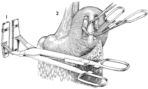
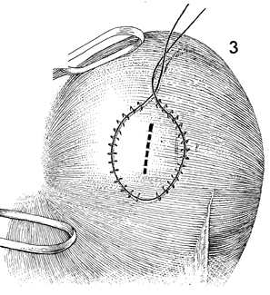
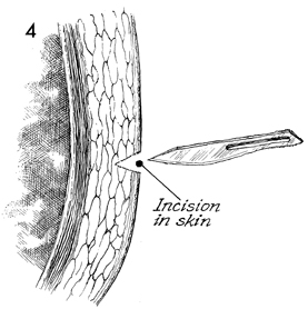
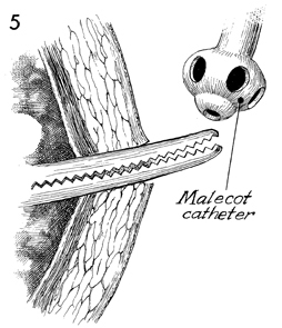
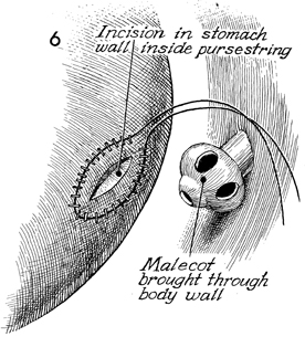
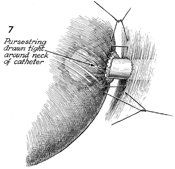
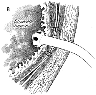
Tube Gastrostomy

A tube gastrostomy can be used following extensive gastrointestinal surgery to decompress the intestines and, when indicated, to supply enteral nutrition.

Physiologic Changes. The gastrostomy tube decompresses the stomach and, while the adynamic process is in place, prevents air from passing into the small bowel. Hydrochloric acid is removed from the stomach. The volume of secretions removed from the stomach should be replaced intravenously with sodium chloride. The alternative to tube gastrostomy is a nasogastric tube. This is a space-occupying mass in the mediastinum. The tube gastrostomy eliminates the need for a nasogastric tube, thereby reducing dead space and  improving respiratory function.

Points of Caution. Care must be taken to see that the gastrostomy tube is within the lumen of the stomach and has not been pulled back into the peritoneal cavity. This is accomplished by suturing the parietal peritoneum to the visceral peritoneum surrounding the gastrostomy and placing skin sutures to the gastrostomy tube.

Technique

This automatic pursestring suture device (United States Surgical Corp.) speeds the placement of the feeding gastrostomy.

The stomach wall is picked up with a Babcock clamp. The automatic pursestring instrument is applied to a zone of the stomach.

An incision is made within the circle of staples placed with the automatic pursestring suture device.

An incision is made on the anterior abdominal wall at a convenient point adjacent to where the stomach would come up to the anterior abdominal wall.

A Kelly clamp is inserted through that incision, and a Malecot catheter is grasped in the Kelly clamp.

An incision is made through the middle of the pursestring sutures into the stomach wall. The Malecot catheter is seen piercing the abdominal wall into the peritoneal cavity.

The tip of the Malecot catheter is pushed through the defect in the stomach. The pursestring suture is tied. Additional sutures are placed between the parietal and visceral peritoneum to seal off the stomach, where the catheter goes through both structures.

The Malecot catheter is seen piercing the abdominal wall, piercing the stomach, and resting within the stomach.

Copyright - all rights reserved / Clifford R. Wheeless, Jr., M.D. and Marcella L. Roenneburg, M.D.
All contents of this web site are copywrite protected.