Home / Site Map / Vulva and Introitus / Vagina and Urethra / Bladder and Ureter / Cervix / Uterus
Fallopian Tubes and Ovaries / Colon / Small Bowel / Abdominal Wall / Malignant Disease: Special Procedures

Malignant Disease:
Special Procedures

Staging of Gynecologic
Oncology Patients With
Exploratory Laparotomy

Subclavian Port-A-Cath 

Peritoneal Port-A-Cath

Application of Vaginal
Cylinders for Intracavitary
Radiation Therapy

Application of Uterine Afterloading Applicators
for Intracavitary Radiation Therapy  

Pelvic High-Dose Afterloader

Abdominal Injection of Chromic Phosphate
()

Supracolic Total Omentectomy

Omental Pedicle "J" Flap

Tube Gastrostomy

Total Vaginectomy

Radical Vulvectomy
With Bilateral Inguinal
Lymph Node Dissection

Reconstruction of the
Vulva With Gracilis Myocutaneous Flaps

Transverse Rectus
Abdominis Myocutaneous
Flap and Vertical Rectus
Abdominis Myocutaneous
Flap

Radical Wertheim
Hysterectomy With
Bilateral Pelvic Lymph
Node Dissection and With Extension of the Vagina

Anterior Exenteration

Posterior Exenteration

Total Pelvic Exenteration

Colonic "J" Pouch Rectal
Reservoir

Kock Pouch Continent Urostomy

Omental "J" Flap Neovagina

Ileocolic Continent Urostomy (Miami Pouch)

Construction of Neoanus
Gracilis Dynamic Anal
Myoplasty

Skin-Stretching System Versus Skin Grafting

Gastric Pelvic Flap for
Augmentation of Continent Urostomy or Neovagina

Control of Hemorrhage in Gynecologic Surgery

Repair of the Punctured
Vena Cava

Ligation of a Lacerated
Internal Iliac Vein and
Suturing of a Lacerated Common Iliac Artery

Hemorrhage Control in
Sacrospinous Ligament
Suspension of the Vagina

Presacral Space
Hemorrhage Control

What Not to Do in Case of Pelvic Hemorrhage

Packing for Hemorrhage
Control

Control of Hemorrhage
Associated With Abdominal Pregnancy

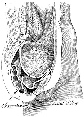
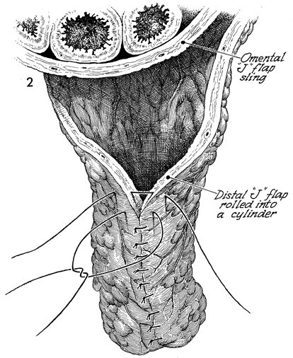
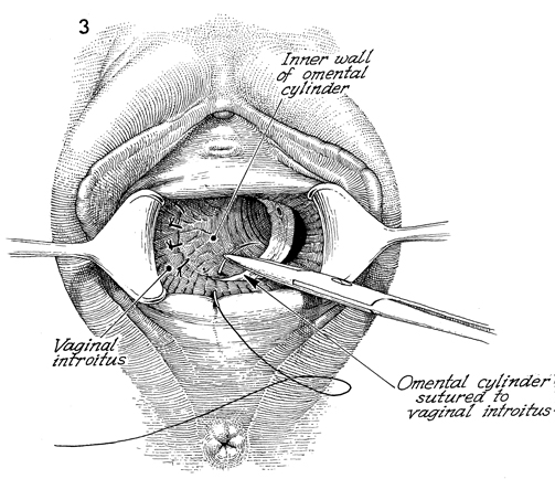
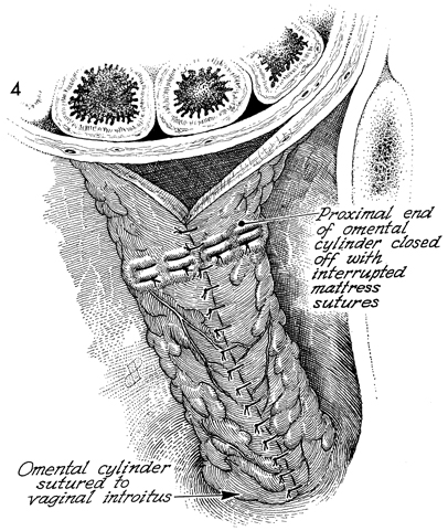
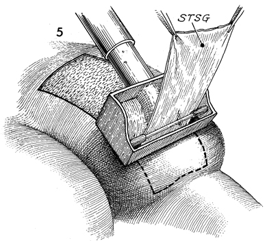
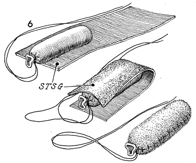
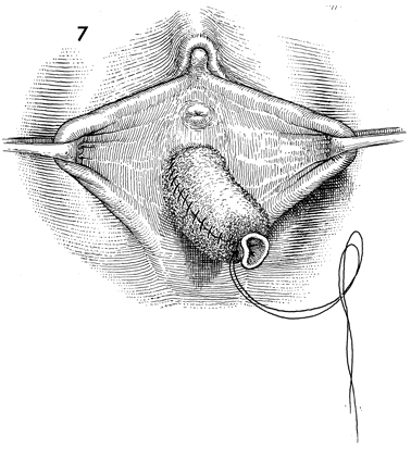
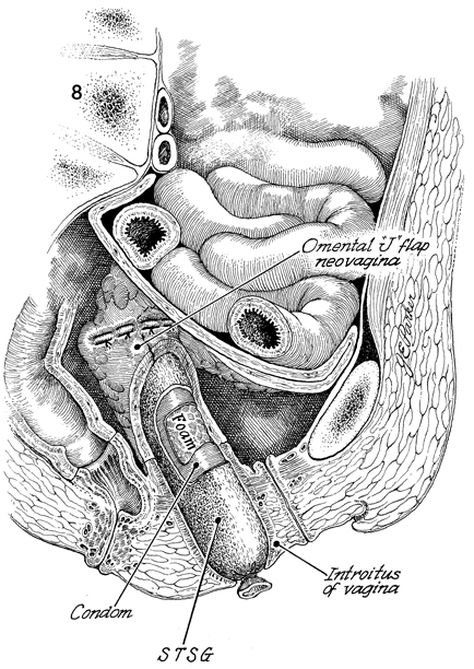
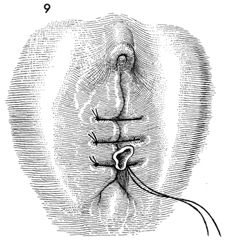
Omental "J" Flap Neovagina

Construction of a neovagina has been an accepted gynecologic procedure for many years. McIndoe (see Vagina and Urethra) has described surgical techniques for construction of a neovagina when the bladder is in position anteriorly and the rectum and colon are in position posteriorly. These techniques are inapplicable, however, when patients have undergone total pelvic exenteration with or without low coloproctostomy. Under these circumstances, Berek and Hacker demonstrated that the anterior wall of the neovagina could be made from an omental flap.

By modifying the omental flap, which is normally used to close off the pelvic inlet after total pelvic exenteration with or without low coloproctostomy, the surgeon can create a cylinder providing anterior, posterior, and lateral walls for the neovagina. When the cylinder is sutured to the introitus and lined with a skin graft, it becomes a satisfactory functional vagina.

Physiologic Changes. The omentum that is enervated by the vagus nerve forms the wall of the neovagina. Normally, tugging or pulling on the omentum does not produce a sensation of pleasure that one would associate with sexual intercourse. Approximately 40% of the patients who have undergone this procedure, however, report that they experience sexual orgasm.

Another physiologic change is the development of estrogen hormone receptors on the split-thickness skin graft. Derived from skin on the buttocks or thigh that normally has no hormonal properties, the graft eventually becomes indistinguishable from normal vaginal mucosa on biopsy. At present, it is unknown whether the maturation index of the graft can be influenced by the administration of systemic estrogen, as can occur in normal vaginal mucosa.

Points of Caution. If the construction of the neovagina immediately follows total pelvic exenteration, it is important to ensure hemostasis in the pelvic wound before applying the skin graft. If hemostasis is uncertain, the omental neovagina should be packed with gauze or foam rubber covered by a contraceptive condom. Then, when hemostasis is maintained, in approximately 6-12 postoperative days a skin graft can be taken and applied to the vaginal form.

After the skin graft has been inserted, the neovagina must remain dilated with a vaginal form until healing is complete. Thereafter, a soft Silastic vaginal form should be worn for 6 months except during intercourse. After this time, the soft Silastic vaginal form is worn only at night if sexual intercourse is not a part of the patient's life.

Technique

This sagittal view shows a patient who has undergone a total pelvic exenteration. In this patient, the rectal stump was left, and the descending colon was brought down for a very low coloproctostomy. The urethra and vagina below the levator sling remain in place. The omentum has been brought down as a flap and has been sutured to the sacral promontory posteriorly and the pubic symphysis anteriorly.

In the upper part of this figure can be seen the omental flap with the intestines lying in the pelvic lid sling. In the lower part of this figure can be seen the distal portion of the flap rolled into a cylinder. The lateral wall of the cylinder has been sutured with interrupted 3-0 polyglycolic acid (PGA) sutures.

This perineal view shows the vulva and vaginal introitus. The wall of the omental cylinder has been sutured to the vaginal introitus with interrupted 3-0 PGA sutures.

The omental cylinder has been completed and sutured to the vaginal introitus.

The dermatome can be seen in this view. STSG, split-thickness skin graft.

The graft is laid out, and a vaginal form is fashioned from foam rubber stuffed into a contraceptive latex condom. The vaginal form has been shaped to an appropriate size, length, and diameter. This is laid on the graft; the graft is folded over the vaginal form, and the edges of the graft are sutured with interrupted 4-0 PGA sutures.

The graft-covered form is inserted through the vaginal introitus into the omental cylinder.

This sagittal section shows the omental "J" flap as the pelvic lid and the residue of the omental flap that forms the outer walls of the neovagina. The graft-covered vaginal form has been introduced into the neovagina.

The labia majora have been approximated loosely by several 2-0 nylon sutures. These remain in place for 10 days. The stump of the condom covering the vaginal form protrudes through the suture line.

On the tenth postoperative day, the patient is returned to the operating room for an examination under anesthesia. The vulvar sutures and vaginal form are removed, and the graft covering the neovagina is inspected.

The patient is fitted with a soft Silastic vaginal form that must be worn for approximately 6 months.

The vaginal form is removed each day and washed, the neovagina is douched, and the form is replaced. Failure to be sexually active and/or to use the vaginal form as prescribed will result in contracture of the neovagina.


Copyright - all rights reserved / Clifford R. Wheeless, Jr., M.D. and Marcella L. Roenneburg, M.D.
All contents of this web site are copywrite protected.