Home / Site Map / Vulva and Introitus / Vagina and Urethra / Bladder and Ureter / Cervix / Uterus
Fallopian Tubes and Ovaries / Colon / Small Bowel / Abdominal Wall / Malignant Disease: Special Procedures

Malignant Disease:
Special Procedures

Staging of Gynecologic
Oncology Patients With
Exploratory Laparotomy

Subclavian Port-A-Cath 

Peritoneal Port-A-Cath

Application of Vaginal
Cylinders for Intracavitary
Radiation Therapy

Application of Uterine Afterloading Applicators
for Intracavitary Radiation Therapy  

Pelvic High-Dose Afterloader

Abdominal Injection of Chromic Phosphate
()

Supracolic Total Omentectomy

Omental Pedicle "J" Flap

Tube Gastrostomy

Total Vaginectomy

Radical Vulvectomy
With Bilateral Inguinal
Lymph Node Dissection

Reconstruction of the
Vulva With Gracilis Myocutaneous Flaps

Transverse Rectus
Abdominis Myocutaneous
Flap and Vertical Rectus
Abdominis Myocutaneous
Flap

Radical Wertheim
Hysterectomy With
Bilateral Pelvic Lymph
Node Dissection and With Extension of the Vagina

Anterior Exenteration

Posterior Exenteration

Total Pelvic Exenteration

Colonic "J" Pouch Rectal
Reservoir

Kock Pouch Continent Urostomy

Omental "J" Flap Neovagina

Ileocolic Continent Urostomy (Miami Pouch)

Construction of Neoanus
Gracilis Dynamic Anal
Myoplasty

Skin-Stretching System Versus Skin Grafting

Gastric Pelvic Flap for
Augmentation of Continent Urostomy or Neovagina

Control of Hemorrhage in Gynecologic Surgery

Repair of the Punctured
Vena Cava

Ligation of a Lacerated
Internal Iliac Vein and
Suturing of a Lacerated Common Iliac Artery

Hemorrhage Control in
Sacrospinous Ligament
Suspension of the Vagina

Presacral Space
Hemorrhage Control

What Not to Do in Case of Pelvic Hemorrhage

Packing for Hemorrhage
Control

Control of Hemorrhage
Associated With Abdominal Pregnancy

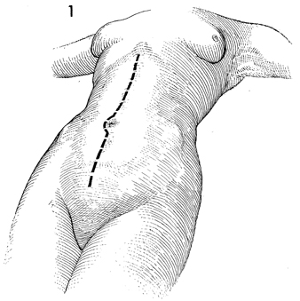
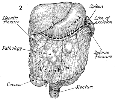
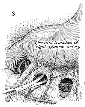
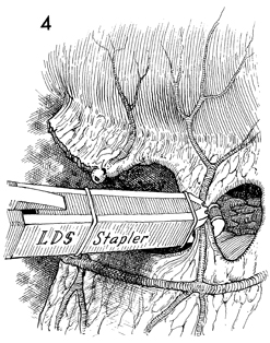
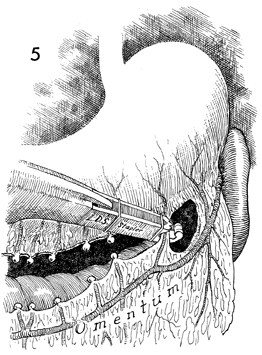
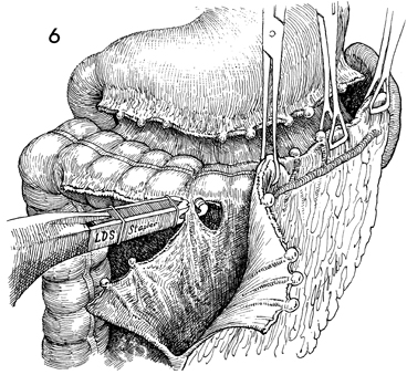
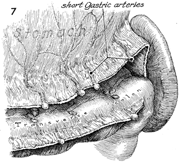
Supracolic Total Omentectomy

Supracolic total omentectomy is performed in conjunction with surgery for ovarian carcinoma. It is important that patients with ovarian carcinoma be operated on through an extended midline incision, generally one from the xiphoid to the symphysis. It is difficult to perform an adequate omentectomy through a transverse or Pfannenstiel incision, and all too often such omentectomies result in incomplete excision of the tumor-bearing omentum, leaving tumor in the remaining portion of the omentum. It is instructive to discover on pathology the degree of micrometastasis in the omentum associated with ovarian carcinoma when clinically the omentum appears to be tumor free.

The purpose of the operation is to remove the total omentum and all gross and microscopic metastases therein.

Physiologic Changes.  None.

Points of Caution.  The omentum should be removed from the greater curvature of the stomach and transverse colon. Care must be taken to secure the small omental branches of the right gastric artery. Meticulous hemostasis should be achieved.

Technique

The incision for total omentectomy must allow exposure to the upper abdomen. This is very difficult with a Maylard-type incision or any type of lower transverse abdominal incision.

There is possible pathology in the omentum. The dotted line represents the line of excision for a supracolic omentectomy. Key anatomical features are the hepatic flexure of the colon, the spleen and its vascular supply, the splenic flexure of the colon, the cecum, and the rectum.

After removing the omentum from the hepatic flexure of the transverse colon, the right gastroepiploic artery and its short gastric branches are identified. Small defects are made between each of the short gastric branches.

The LDS (linear dissecting) stapler (United States Surgical Corp.) is applied to each of the short gastric branches by manipulating the stapler into the defects created in the omentum.

The omentum is completely removed from the stomach.

The left gastroepiploic artery has been stapled. The remaining omentum is removed from the transverse colon.

The stomach and short gastric arteries are noted. The transverse colon has been cleaned, and the omentum totally removed.


Copyright - all rights reserved / Clifford R. Wheeless, Jr., M.D. and Marcella L. Roenneburg, M.D.
All contents of this web site are copywrite protected.