Home / Site Map / Vulva and Introitus / Vagina and Urethra / Bladder and Ureter / Cervix / Uterus
Fallopian Tubes and Ovaries / Colon / Small Bowel / Abdominal Wall / Malignant Disease: Special Procedures

Vagina and Urethra

Anterior Repair and Kelly Plication
           
Site Specific Posterior Repair
           
Sacrospinous Ligament Suspension of the Vagina
           
Vaginal Repair of Enterocele
           
Vaginal Evisceration
           
Excision of Transverse Vaginal Septum
           
Correction of Double-Barreled Vagina
           
Incision and Drainage of Pelvic Abscess via the Vaginal Route
           
Sacral Colpoplexy
           
Le Fort Operation
           
Vesicovaginal Fistula Repair
           
Transposition of Island Skin Flap for Repair of Vesicovaginal Fistula
           
McIndoe Vaginoplasty for Neovagina
           
Rectovaginal Fistula Repair
           
Reconstruction of the Urethra
           
Marsupialization of a Suburethral Diverticulum by the Spence Operation
           
Suburethral Diverticulum via the Double-Breasted Closure Technique           
           
Urethrovaginal Fistula Repair via the Double-Breasted Closure Technique
           
Goebell-Stoeckel Fascia Lata Sling Operation for Urinary Incontinence
           
Transection of Goebell-Stoeckel Fascia Strap
           
Rectovaginal Fistula Repair via Musset-Poitout-Noble Perineotomy

Sigmoid Neovagina

Watkins Interposition Operation

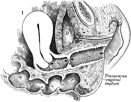
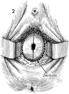
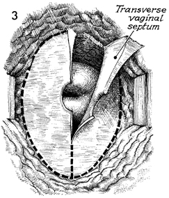
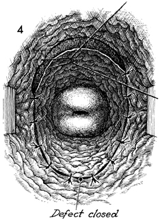
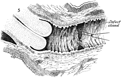
Excision of Transverse Vaginal Septum

Transverse vaginal septum generally occurs between the upper one-third and lower two-thirds of the vaginal canal. This is additional evidence that the upper one-third of the vagina is of Mullerian origin and that the lower two-thirds of the vagina is embryologically developed via the urogenital sinus.

The septum can be either complete or partial. If it is complete, the symptoms of vaginal obstruction occur at the time of menarche, since menstrual blood is entrapped above the septum and has no egress from the vagina. If the septum is partial, it may be discovered on a routine gynecologic examination, or the patient may present with dyspareunia.

The operation is performed to remove the transverse vaginal septum without significantly shortening the vaginal canal.

Physiologic Changes. The physiologic changes desired are (1) the egress of menstrual blood from the vaginal canal without obstruction and (2) the normal functioning of the vagina.

Points of Caution. If the septum is complete and hematometra or hematocolpos is present, it is unwise to attempt surgical correction of the septum at the time the obstruction is relieved. The procedure of choice is incision and drainage of the hematometra or hematocolpos, with reconstruction delayed 6-8 weeks until the tissues have completely healed.

To avoid unduly shortening the vagina, excessive vaginal mucosa should not be removed.

Technique

The typical position of most transverse vaginal septa at the junction of the upper one-third and the lower two-thirds of the vagina is shown. B, bladder.


With the patient in the dorsal lithotomy position, the perineum is prepped and draped, and adequate vaginal retraction is applied to allow exposure of the septum, which is incomplete here. Initially, the septum is grasped with Allis clamps, and a vertical incision is made through the septum to divide it in half.


The septum is picked up with tissue forceps, traction is applied, and with a scalpel the septum is separated from the vaginal mucosa.

The vaginal mucosa is then approximated with interrupted 3-0 synthetic absorbable suture throughout its circumference.

A sagittal view illustrates closure of the defect in the vaginal mucosa.


Copyright - all rights reserved / Clifford R. Wheeless, Jr., M.D. and Marcella L. Roenneburg, M.D.
All contents of this web site are copywrite protected.