Home / Site Map / Vulva and Introitus / Vagina and Urethra / Bladder and Ureter / Cervix / Uterus
Fallopian Tubes and Ovaries / Colon / Small Bowel / Abdominal Wall / Malignant Disease: Special Procedures

Abdominal Wall

Pfannenstiel Incision

Maylard Incision

Panniculectomy

Incisional Hernia
Repair

Abdominal Wound
Dehiscence and
Evisceration

Massive Closure
of the Abdominal
Wall With a One-Knot
Loop Suture

Hemorrhage Control Following Laceration
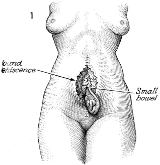
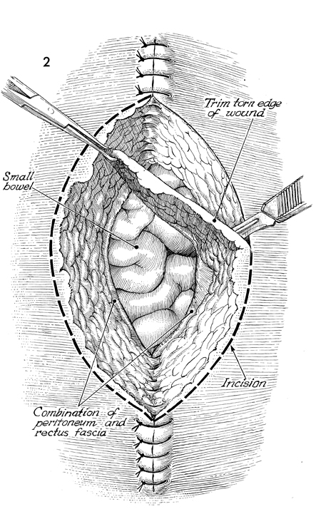
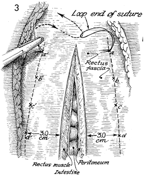
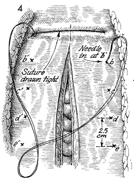
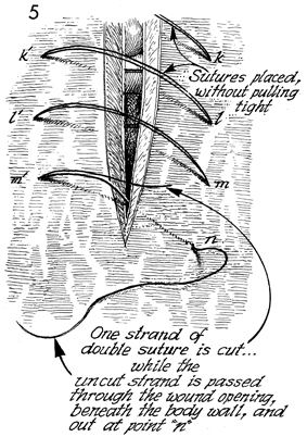
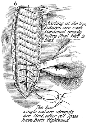
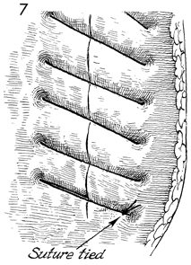
of Inferior Upper
Epigastric Vessels

Abdominal Wound Dehiscence
and Evisceration

The seepage of serosanguineous fluid through a closed abdominal wound is an early sign of abdominal wound dehiscence with possible evisceration. When this occurs, the surgeon should remove one or two sutures in the skin and explore the wound manually, using a sterile glove. If there is separation of the rectus fascia, the patient should be taken to the operating room for primary closure. Wound dehiscence may or may not be associated with intestinal evisceration. When the latter complication is present, the mortality rate is dramatically increased and may reach 30%.

The basic principles of management of abdominal wall dehiscence and evisceration are early diagnosis and surgical closure. The latter is accompanied by mass closure with wide sutures of heavy delayed synthetic absorbable suture.

The purpose of the operation is to close the abdominal wall.

Physiologic Changes. Dehiscence may stem from wound hematomas or from excessive intra-abdominal pressure secondary to abdominal coughing or vomiting that has disrupted the sutures. It is most commonly seen in patients with properties of poor wound healing, such as patients with diabetes, oncology patients, and patients taking steroid medications.

Points of Caution. All attempts should be made to diagnose and manage this problem promptly to minimize the risk of intestinal evisceration.

All sutures should be placed prior to tying any one suture.

Technique

The patient showing abdominal wall dehiscence with evisceration of the small intestine is placed in the supine position under general anesthesia.

The contaminated edges of the wound including a combination of the peritoneum and rectus fascia are excised.

At the xiphoid end of the incision, the needle is placed through point a to point a' through all layers of rectus fascia muscle and peritoneum. When the needle is brought out of point a', it is passed through the loop end of the suture.

The needle is brought through point b to point b', 2 1/2 cm from point a and point a', respectively.

The sutures are not pulled taut for the remainder of the suture c through n, m, etc. By leaving the sutures loose, the surgeon has the best opportunity to place the sutures precisely. When the sutures have been completely placed, they can be cinched up taut. Here one sees the technique of placing the final sutures from m' to n. One strand of the double suture is cut, while the uncut strand is passed through the wound opening beneath the abdominal wall and out of point n from inside to out.

The sutures are cinched up snugly but are not tight. The two single-suture strands are tied after all the loops have been tightened. Multiple throughs, more than five, are placed in this knot.

The suture has been tied. To date, there has been no item of data to show that this closure has any less or any greater strength than the more time-consuming Smead-Jones closure with running far-to-near-near-far suture.


Copyright - all rights reserved / Clifford R. Wheeless, Jr., M.D. and Marcella L. Roenneburg, M.D.
All contents of this web site are copywrite protected.