Home / Site Map / Vulva and Introitus / Vagina and Urethra / Bladder and Ureter / Cervix / Uterus
Fallopian Tubes and Ovaries / Colon / Small Bowel / Abdominal Wall / Malignant Disease: Special Procedures

Fallopian Tubes
and Ovaries

Laparoscopy Technique

Diagnostic Uses
of Laparoscopy

Demonstration
of Tubal Patency
via Laparoscopy

Laparoscopic Resection
of Unruptured
Ectopic Pregnancy

Ovarian Biopsy
via Laparoscopy

Electrocoagulation of
Endometriosis via
Laparoscopy

Lysis or Adhesions
via Laparoscopy

Control of Hemorrhage
During Laparoscopy

Fallopian Tube
Sterilization

Sterilization by
Electrocoagulation and
Division via Laparoscopy

Silastic Band Sterilization
via Laparoscopy

Hulka Clip Sterilization
via Laparoscopy

Sterilization by the
Pomeroy Operation

Sterilization by the
Modified Irving Technique

Sterilization by the
Minilaparotomy Technique

Sterilization - Ucheda Technique

Salpingectomy

Salpingo-oophorectomy

Fimbrioplasy

Tuboplasty -
Microresection
and Anastomosis
of the Fallopian Tube

Wedge Resection
of the Ovary

Torsion of the Ovary

Ovarian Cystectomy

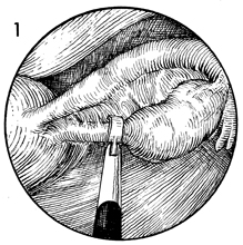
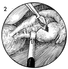
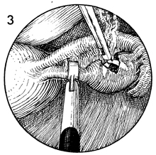
Ovarian Biopsy via Laparoscopy

Biopsy of the ovary is rarely necessary. Modern cytogenetic and endocrine laboratory techniques can usually ascertain whether the ovary contains oocytes. There are some cases, however, in which biopsy of the ovary may be indicated.

The purpose of the operation is to obtain an adequate biopsy of the ovary through the laparoscope.

Physiologic Changes. Removal of a piece of ovary can, in some cases, change the physiology of the hypothalamic-pituitary-ovarian axis in the same manner as wedge resection of the ovary alters the physiology in polycystic ovary disease.

Points of Caution.  The predominant complication from ovarian biopsy is control of hemorrhage from the bed of the ovary. Thorough electrocoagulation of the entire biopsy site should be performed. The site should be observed for at least 3-4 minutes to ensure that hemostasis is complete.

Technique

Ovarian biopsy by laparoscopy requires a two-incision technique. It is preferable to use an operating laparoscope with a 3-mm grasping forceps to grasp the suspensory ligament of the ovary and anchor the ovary in a stable position.

A 6-mm alligator biopsy forceps is passed through a second-incision trocar, and a large bite of ovarian capsule and stroma is taken. Bleeding from this site can be copious and must be coagulated. If bleeding obscures observation of the biopsy site, a third puncture is made in the abdominal wall. A 2-mm aspiration needle connected to a 50-mL syringe of saline solution is introduced through the puncture, and the biopsy site is irrigated with saline solution rather than suctioned.

A large electrocoagulation biopsy forceps is inserted through the second-incision trocar into the biopsy wound, and the jaws of the forceps are opened. The electrical current is applied, and the ovary is coagulated thoroughly from the inside. This usually stops most of the bleeding. The site is irrigated with saline solution. The biopsy forceps can be applied to small bleeding areas with the jaws in the closed position, providing point cautery rather than widespread cautery. The ovary should be observed for at least 3-4 minutes to ensure that all bleeding is stopped prior to removing the instruments from the abdomen.


Copyright - all rights reserved / Clifford R. Wheeless, Jr., M.D. and Marcella L. Roenneburg, M.D.
All contents of this web site are copywrite protected.